Explanations of terms and possible solutions
Explanation of the term spine
... all about spinal problems
The spine forms the supporting organ for the upper body and houses the central nerve cord (myelon). The intervertebral discs are responsible for the mobility of the spine and individual sections. Damage to the spine can therefore cause severe symptoms, often affecting organs and limbs that are not directly affected. Most of these symptoms are caused by muscle tension or wear and tear on the intervertebral disc.
Problems with the spine are one of the most common reasons for sick leave in Germany. More than 80% of the population have back problems at least once in their lives.
The APEX Spine Center has set itself the task of helping to make the variety of terms relating to the spine easier to understand for you as a patient and to provide you with a comprehensible guide so that you can better classify and understand the medical terms for yourself . At the same time, it should then be easier for you to find the optimal and gentlest solution to your problem.
 Cause of pain:

The causes of back pain are varied. On the one hand, the complaints/pain can come from the front area of the spine (intervertebral disc, vertebrae) and/or from the rear area of the spine (bone changes due to facet arthrosis, stenosis, instability, etc.). The most common are degenerative changes in the spine (about 80%). Of course, diseases such as rheumatism, traumatic changes (fracture of the vertebral body), inflammatory diseases and tumors are also treated in the APEX Spine Center.
General cause of pain
- Musculature (e.g. due to overload or tension)
- intervertebral disc
- vertebral joints
- Main nerve and nerve branches (outlets)
Causes of spinal pain
degenerative changes
- intervertebral disc changes
- ligamentous insufficiency (= functional disorders of retaining ligaments)
- bony changes
- Disturbance of the articulated vertebral connections
deformities
- Growth disorders (e.g. scoliosis)
- Congenital malformations
 Generalized skeletal disorders
- osteoporosis
- Osteomalacia (= softening of the bones)
Inflammatory diseases
- infectious diseases
- Rheumatic forms
tumors of the spine
- metastases
- primary tumors
Dream
- fractures
- whiplash of the lumbar spine
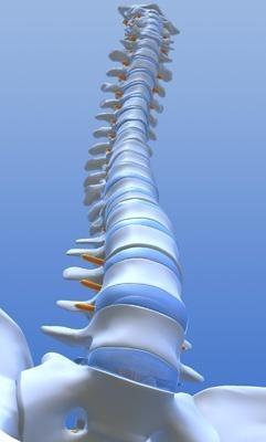
Different regions of the spine
Four main areas are distinguished
- the cervical spine, consisting of 7 vertebral bodies (also called the cervical spine section);
- the thoracic spine, consisting of 12 vertebral bodies (also called the thoracic or thoracic region);
- the lumbar spine, consisting (with a few exceptions) of 5 vertebral bodies (also called LWS - or lumbar area)
- and the fourth area, the sacrum (Os sacrum) and the coccyx (Os coccygeum), which generally has less need for therapy.
Each area of the spine has an individual accumulation of different clinical pictures due to the anatomy (body plan) and the "disease-causing" typical stresses in today's modern working world, for which in turn a large number of different terms are used.
leading symptoms
The main symptom of a herniated disc is pain.
Sometimes accompanied by a pain- and static-related malposition and a paresthesia (numbness) that can be assigned to the affected root.
Serious symptoms (signs of illness) that often require immediate action are:
- paresis (paralysis)
- Bladder and defecation disorders
- sudden loss of sexual function
The reasons for this are often wear and tear (degeneration) of the intervertebral discs with the following loss of height (:= osteochondrosis) and continuous overstretching of the joint capsules and the development of accompanying arthrosis of the small vertebral joints (:= facet arthrosis). As a reaction to this, the muscles in the immediate vicinity tense up. Especially in the area of the spine, the so-called paravertebral muscles (the accompanying rear muscle strands to the right and left of the axis organ) tend to weaken, tighten and shorten (:= hypotrophy, induration). The front so-called oblique abdominal muscles tend to weaken under constant spinal pain - up to the point of disappearing completely (:= atrophy).
Different types of pain:
The pain, in turn, can be local, narrowly defined or punctiform.
It can have an extensive character (usually in the case of extensive findings associated with reactive, extensive muscle tension).
It can be pseudoradicular (pain that is transmitted via so-called muscle chains) radiating into the arm - or the leg, but without an explanatory cause being found during the neurological examination. This type of pain is usually of a dull character and can often only be vaguely explained by the patient be specified.
It can also have a radicular character. Here, clear damage to the nerve substance is identified as the cause of the symptoms. The character of the pain is pointed, sharp and its extent is narrowly circumscribed and clearly attributable to the so-called dermatomes (areas of skin that can be assigned to the nerves).
Blockages in the spine
Vertebral joint blockages are a special form of spinal complaints.
The blockade describes a (similar to a jammed drawer) extensive inability of the individual joint partners to move or slide against each other in the anatomically specified area.
This temporary suspension of the joint function of the facets (= vertebral joints) can overtake a young patient without wear and tear symptoms as well as an older patient with pronounced wear and tear symptoms. Reasons for the development of a blockage are local overloads due to unusual physical and sporting exertion. Sometimes the component of hypothermia is added to this. Some blockages are caused by spinal compression injuries,
In most cases, simple blockages can be remedied promptly with chiropractic or manual therapy treatments.
Manipulations (through a small, non-violent movement impulse) or mobilizations (through gentle stretching of the joints, muscles and connective tissue structures in the restricted direction) are used as treatment measures. The release of the blockage is sometimes accompanied by a "cracking". More stubborn blockages must be treated in multiple sessions.
Chiropractic must be distinguished from manual therapy / chirotherapy. The chiropractors are non-medical professionals and do not treat according to the specified guidelines for manual therapy (which apply throughout Europe).
However, a blockage, if it is diagnosed as such, can always be an expression of previously undiscovered intervertebral disc symptoms - or stenosis symptoms. In this case, the doctor treating you must pay particular attention to the physical examination, the questioning of the patient (anamnesis) and the diagnosis (if possible, carrying out a high-resolution nuclear spin tomogram).
Frequency of affected individual sections of the spine
The region most frequently affected by symptoms is the lumbar spine (lumbar spine), most of which are the two lowest intervertebral disc levels (L5/S1 and L4/5),
followed by the cervical spine (cervical spine) and finally the thoracic spine (thoracic spine).
Explanation of terms related to spinal disorders
General definition of terms
Due to the lack of exercise caused by the "modern way of life" and the resulting weakening of the trunk muscles, coupled with wear and tear of the intervertebral discs and the vertebral joints, which lead to stenosis problems in the case of intensive development, a multitude of expressions are used colloquially and also in medical practice, which are explained below should be.
Back pain in the lumbar spine area is often referred to as "the intervertebral disc" or "lumbago" ( = local back pain often associated with a significant restriction of movement, a possible vertebral joint blockage and accompanying muscle tension).
A distinction is made between the so-called sciatica (also lumboischialgia or sciatica, corresponding to whether the pain extends from the back into the leg - or is only present as leg pain).
The term spinal wear includes the degenerative change in the intervertebral disc space with the loss of the elastic buffering effect of the intervertebral disc and the resulting changed base and cover plates with the x-ray-typical bone extensions (the so-called spondylosis). If there is also local muscle tension that is difficult to control with edema formation (water retention) in the vertebral body base and cover plate areas, which can often be seen in the MRI image, this is referred to as activated osteochondrosis.
disc problems
If the symptoms come from the intervertebral disc (synonym: discus or discus intervertebralis) itself (:= discogenic cause), then many different expressions are often used for the respective pathological condition of the intervertebral disc itself.
The shape of the intervertebral disc consists of a fibrous ring (:= anulus fibrosus) and a gelatinous core (the nucleus pulposus). The fibrous ring forms the outer mechanical shell and the inner gelatinous core provides the necessary elastic properties (buffer effect).
One speaks of a discopathy when there is a general pathological disorder of the intervertebral disc function. This usually begins with a loss of fluid from the
gelatinous core and a corresponding reduction in height of the intervertebral space (synonym: intervertebral space) This condition of the reduced space between the adjacent vertebral bodies is called osteochondrosis.
The shifting of the intervertebral disc material backwards in the direction of the spinal cord and/or also in the direction of the nerve root, which occurs parallel to this condition, is described with different terms.
In the event of disc damage, the fibrous ring (anulus fibrosus) is affected first, in that it partially or completely tears (so-called annulus rupture). The result can then be a more or less pronounced displacement of the gelatinous core (nucleus pulposus).
If the intervertebral disc slips only slightly from the resulting tear in the fibrous ring
(annulus rupture) herraus, one speaks of a disc protrusion or a protrusion. Due to its severity, this is divided into protrusions (bulging discs) of the first and second degree. However, all of these protrusions have in common that the fibrous ring (anulus fibrosus) is not complete
is torn through, i.e. with certain parts the ring still holds together in its form.
Consequently, in the event of a herniated disc (synonyms: prolapse, nucleus pulposus hernia, nucleus pulposus prolapse), the fibrous ring (anulus fibrosus) is completely torn and the disc material (= the gelatinous core) enters the front part of the spinal canal
(the intervertebral disc “slips out”) and leads to nerve compression there.
A special feature of the herniated disc is the sequester, or the sequestered herniated disc. Here the intervertebral disc (the gelatinous core) slips completely into the spinal canal and loses the connection to the intervertebral disc space.
The resulting therapy can be an intervertebral disk operation (synonyms: nucleotomy or discotomy) if the conservative attempt is unsuccessful.
(The term discotomy rather imprecisely describes the process of the operation, since essentially the nucleus pulposus = gelatinous core is removed to the part that presses on the nerve substance and the fibrous ring itself remains untouched)
→ The particularly gentle form of therapy for surgical disc treatment is endoscopic nucleotomy, which we have been successfully performing for many years. Unlike other surgeons, we can say that any herniated disc can be treated with this method. (see also scientific publications)
The vertebral arthrosis
If the wear-changed small, paired vertebral joints
(:= facets) cause symptoms, one speaks of spondylarthrosis, facet arthrosis, a facet syndrome, vertebral joint arthrosis or arthrosis of the interarticular portion.
In the advanced stage, these vertebral joint arthrosis are the most common causes for the development of a spinal canal stenosis (:= spinal canal stenosis, spinal stenosis, narrowing of the spinal canal, narrowing of the spinal canal).
The spinal canal stenosis
In a pronounced form, in which many movement segments (definition according to Junghans: a pair of vertebral bodies with all jointly adjacent structures) are affected by spinal canal stenosis (spinal canal stenosis), one speaks of MESS, the multi-segmental bottleneck syndrome of the spinal canal.
Other causes of this quite common but often unrecognized clinical picture are:
- Sporting and professional overload over many years as well
- the congenital narrowing of the spinal canal (-> synonym: congenital stenosis).
The diagnosis of spinal canal stenosis can never be made solely by measuring the horizontal (transversal) nuclear spin slices alone, but it must always match the symptoms.
At this point it should be mentioned that the width, ie the extent of the spinal cord, varies from patient to patient, and the sensitivity of the spinal cord membrane (:= dura) varies from person to person.
All of this makes the correct diagnosis a sometimes demanding endeavor that requires a certain clinical experience of the doctor.
Therapy of the stenosis:
Therapy of advanced stenosis with claudication and spinal symptoms
(Stenosis-related “window sickness”, the clearly painful restriction of walking distance) is an operative domain. Conservative measures such as injections, physiotherapy and electrical treatment usually fail miserably.
Thanks to the tissue-sparing surgical technique, we are able to
( Microscopic decompression of the spinal canal ) to release the patient a few days after the operation and, after a short outpatient rehabilitation, to integrate him back into normal life as quickly as possible.
The Sliding Swirl
A special form of stenosis is the "slipped vertebra", the so-called sliding vertebrae ( spondylolisthesis or listhesis ). This form of spinal disease is also optionally associated with a more or less severe form of segmental instability. This means that the affected vertebrae tend to slide away from one another transversely (→ from back to front) under the appropriate load.
A distinction is made between spondylolisthesis vera (the "real", the "true" sliding vertebrae), which is associated with a congenital abnormal adhesion of the vertebral arch (the so-called spondyloylsis).
from pseudospondylolisthesis.
This is usually accompanied by a lower degree of sliding and is based on a congenital misalignment of the vertebral joint position coupled with wear and tear on the vertebral joints.
With regard to the degree of glide, there are a wide variety of classification methods. The classification according to Meyerding (grades I to IV) is still the most common.
The complete slipping of the gliding vertebra forward and down is called spondyloptosis.
Form of therapy: In the case of an indication for surgery, we can use the technique of ( microscopic decompression of the spinal canal ) in many cases by preserving the interarticular portion (intervertebral joints, facets) to a simultaneous transpedicular reposition spondylodesis ( -> synonym: stiffening operation with simultaneous correction of the slipped vertebra ) waive!
The degenerative lumbar scoliosis
This wear-related lateral deflection with simultaneous "twisting" (→ rotation) of the vertebrae at the apex of the scoliosis is caused simultaneously by existing stenoses and the increasing incorrect static.
The development of scoliosis can have different causes:
- degenerative (due to wear)
- caused by a pre-existing infantile, juvenile or congenital scoliosis
- traumatic (caused by an accident)
- metabolic
The (advancing) degenerative lumbar scoliosis is a common cause of degenerative symptoms from the 4th to 5th decade of life.
In addition to purely degenerative reasons, the second most common cause is juvenile (-> originating in adolescence) or infantile (originating in childhood), or congenital (congenital) scoliosis.
Often the scoliosis causes only moderate symptoms through physical compensation (- good trunk muscle corset!) for years. Especially in the lumbar spine area, scoliosis tends to progress from the 4th to 5th decade of life to progressive side bending with a simultaneous, statically induced worsening of the measurable scoliosis angle.
In the case of a scoliosis that is predominantly pronounced in the thoracic spine area, the progression (-> synonym: progression) is much less pronounced due to the external support of the laterally attached scoliosis.
Therapy:
Conventional treatment with electrotherapy, physiotherapy and support garments often brings only minimal relief, if at all.
The cause of degenerative lumbar scoliosis can only be successfully treated by invasive removal of the "bottlenecks" responsible for the symptoms.
The subtle, minimally invasive and tissue-sparing surgical technique
( Microscopic decompression of the spinal canal ) allows us to remove the bony and also ligamentous stenosis in almost all cases without extensive surgical stiffening, naturally protecting the load-bearing facets (vertebral joints).
The "conventional (normal) standard solution" in almost all clinics is often multi-level decompression through laminectomy and subsequent transpedicular distraction spondylodesis using double-rod instrumentation.
We believe that in many cases our patients can be spared this long-distance stiffening operation, with all its disadvantages, and that we can significantly alleviate their symptoms and consequently improve their quality of life.