
Diagnosis
... the be-all and end-all of the treatment concept at the apex spine center
Unfortunately, not only general practitioners but also orthopedic surgeons and neurosurgeons tend all too often to subject the patient to overtreatment without precise knowledge of the diagnosis - i.e. the cause of the pain. In order to be able to make a well-founded diagnosis of your back pain, a conscientious and detailed procedure is essential. This generally requires different preliminary examinations in the sense of step-by-step diagnostics. These include:
- Anamnesis / previous history, knowledge of the medical history forms the basis of a reasonable and serious diagnosis
- Detailed targeted orthopedic-neurosurgical and neurological examination
- Strength test of the lumbar extensors and neck muscles
- electromyographic examination (EMG), spine motion
- Internal examination using ultrasound and blood count diagnostics (infection diagnostics, rheumatism, gout serology
- X-ray examination
- Nuclear spin tomography (magnetic resonance tomography, MRT), provides an exact imaging of all relevant structures affecting the spine (bony spinal canal, condition of the intervertebral disc, representation of the spinal cord and spinal nerves)
Latest MRI technology in the apex spine center!
A 1.5 tesla MRT device of the latest generation has recently been available for you as a patient in our premises. With this new device, the sometimes long examination times are significantly reduced. We can now determine the cause of your neck and back pain even faster.
What advantages do you as a patient have from MRI diagnostics directly at the apex spine? 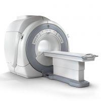
For you as a patient, there are no unnecessary delays and waiting times until all the necessary examinations have been completed.
Your advantage: after your visit to our spine center, you go home with a written report including a diagnosis and a suggested treatment. This saves you longer delays and you know with just one visit where the cause of your problem lies.
What is magnetic resonance imaging?
Magnetic resonance imaging (MRI), also known as nuclear spin tomography, is an imaging method for generating cross-sectional images of the entire body. Due to the high-contrast tissue display, internal structures such as a herniated disc , bony spinal canal stenosis, tumors or foci of infection can be displayed exactly. Magnetic resonance tomography has been used successfully in medical diagnostics worldwide since the mid-1980s. How does magnetic resonance imaging (MRI) work
MRI does not use X-rays, just a strong magnetic field and radio frequency waves - similar to those used by radio stations. Highly sensitive antennas pick up the necessary information from the body. These signals are then converted into sectional images of the body with the help of computers. As a result, the patient is not exposed to X-rays.
If the results are not indicative ( herniated disc , spinal canal stenosis , degeneration or osteochondrosis, inflammation), further diagnostics, such as a discography, may be necessary. Here, a contrast medium is injected into the intervertebral disc using local anesthesia through a very fine needle. Based on the distribution image of the contrast agent and the patient's reaction (e.g. typical pain, memory pain), the diseased intervertebral disc causing the symptoms can be identified ( prosthesis and/or endoscopic surgery ).
If the way to our practice is too far for you, you also have the opportunity to get a preliminary assessment of your complaints by filling out our questionnaire conscientiously. To do this, please follow the following link: Rapid diagnosis and advice