Quick overview herniated disc :
Lumbar spine
Cervical Spine
Herniated disc
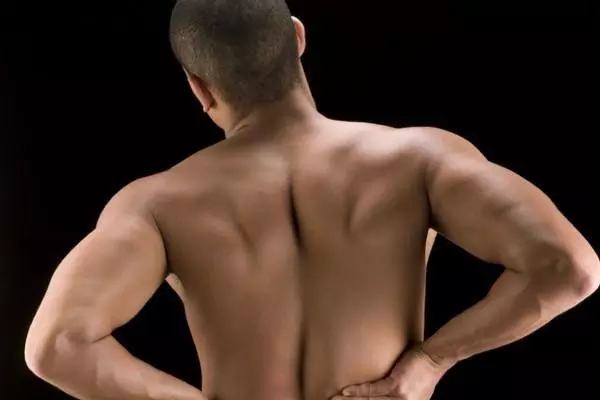
Herniated disc in the area of the lumbar spine
Lower back pain can have many causes. In the case of acute pain, a herniated disc in the lumbar spine can only be expected in about three to five percent of cases. Surgery is not always necessary, since in about 90% of herniated discs the material that protrudes is resorbed over time. A significant improvement in symptoms can be expected after about four to six weeks. If this does not occur and other symptoms (e.g. sensory disturbances) occur, a check should be carried out and an intervertebral disc operation may be indicated.
The following symptoms indicate a herniated disc in the lumbar spine:
- Severe pain in the lower back
- Increased pain in the buttocks, legs and/or feet
- Sneezing, coughing, or straining makes the pain worse
- The pain increases when bending, sitting or lying down
- Sensitivity disorders or signs of paralysis can also occur
- Numbness in the legs or buttocks
- Loss of bladder and rectum control
Not all symptoms are always involved. The pain can also vary in severity. The latter are a sign that nerve involvement is present. Here the extent of the nerve compression or damage determines the severity of the symptoms.
The so-called cauda equina syndrome occurs very rarely. This is an emergency that requires immediate surgical attention to avoid permanent damage. With this symptom, disc material compresses the spinal nerve roots of the "cauda equina" and leads to complex neurological deficits. Paralysis of the legs, sensory disturbances, micturition and defecation disorders as well as a disturbance of the sexual function occur. Surgery must be performed within a few hours of the onset of symptoms, otherwise nerve damage may remain. The goal of every disc surgery is the decompression of the nerves. The faster this happens, the better the prognosis.
What happens when there is a herniated disc in the lumbar spine?
The intervertebral disc is also called the intervertebral disc or discus intervertebralis. Humans have 23 pieces, which can be between 5 and 20 mm thick. They get thicker from the cervical spine, through the thoracic spine to the lumbar spine. The intervertebral discs act as shock absorbers and protect the vertebrae by absorbing and dampening shocks. In addition, they make the spine flexible and create space for the nerve outlets from the nerve exit holes, the so-called foramen. The foramen are formed on both sides by two vertebral bodies and the intervertebral disc, the so-called segment.
The structure of the intervertebral disc consists of an outer fibrous ring (anulus fibrosus) and the inner gelatinous core (nucleus pulposus). If this outer ring of fibers is weakened (e.g. due to degeneration and collagen breakdown), it can crack when subjected to unfavorable movement or high loads, comparable to a porous rubber tire. These tears can become larger and, in extreme cases, disc material from the gelatinous core can protrude or detach completely and then press on the spinal nerves. On the one hand, this causes acute back pain and/or leg pain and, on the other hand, neurological deficits can also occur.
The herniated disc in the lumbar spine is the most frequently occurring herniated disc at well over 90%. It usually occurs in the lumbar region between lumbar vertebrae four and five (L4/L5) or between the last lumbar vertebra and the first sacral vertebra (L5/S1). Herniated discs in the cervical spine are rare. Extremely rare (less than 1/1000) in the area of the or thoracic spine.
What can trigger a herniated disc in the lumbar spine area?
These reasons can lead to a herniated disc in the lumbar spine:
- Weakening of the fibrous ring due to age-related degeneration
- Weakening of the fiber ring due to incorrect and excessive strain (e.g. sitting for a long time and not moving enough)
- Congenital disc weakness
- Poorly developed back muscles in the area of the lumbar spine
- Jerking or twisting movements of the upper body
- Lifting or pushing heavy objects
- Genetic factors such as back problems that run in families or herniated discs
- Unbalanced diet
However, a herniated disc can also occur without a recognizable trigger.
Treatment of a herniated disc
Many herniated discs do not require surgery. In about 80% of all known cases of back pain caused by a herniated disc, conservative treatment measures are successful. Back pain in the area of the lumbar spine can be effectively combated with an individually created therapy plan and additional pain medication. As soon as the symptoms improve, a training program can be started that gently strengthens the abdominal and back muscles.
Conservative therapy for a herniated disc in the lumbar spine
In the case of a herniated disc, the first step in the acute phase is to immobilize and protect the back for two to four days. Painkillers are taken at the same time to suppress the acute pain.
After the acute phase, it’s time to move – move – move! After that, an individual training plan is created in our apex spine center under the guidance of a doctor or with physiotherapeutic advice, which is intended to strengthen the back and abdominal muscles slowly and while protecting the lumbar spine. Electrotherapy, heat treatments, or massage can help build muscle and speed recovery. It is recommended to resume normal activities as soon as possible, as exercise relieves back pain more quickly. Too long closed periods can be counterproductive. In a study by Keel et al. it was shown that out of 100 examined patients with acute back pain in the lumbar region, more than 40% were able to return to work more quickly (after 10 days) if they quickly returned to normal everyday activities. Patients who rested for longer also took longer to recover.
If the pain is very severe, it can also be relieved with the help of injections in the area of the herniated disc or the affected lumbar region. Here, numbing substances are usually injected in combination with cortisone preparations next to the pinched nerve fiber or next to the spinal cord. The cortisone reduces the swelling and thus reduces the pain. In the case of severe neurological symptoms, treatment in a clinic may also be necessary. In severe cases, the pain and cortisone medication can be administered there by means of an infusion.
| Too much protection and rest can damage 1) | ||
|---|---|---|
| Out of 100 patients with acute back pain are: |
patients as possible resume their daily activities early record, tape |
patients who become stronger and spared longer |
| again after 10 days able to work |
57 | 36 |
| after a month still ill |
17 | 31 |
Surgical therapy for a herniated disc in the lumbar spine
Surgical intervention is indicated for severe disc herniation or when symptoms worsen. Open back surgery is performed under general anesthesia in over 98% of spine centers in Europe. The surgeon clears out the protruding disc material to decompress the pinched nerves. Occasionally, an intervertebral disc prosthesis is implanted at the same time, which maintains the mobility of the spine. This is followed by a long rehabilitation phase.
There is now also the option of minimally invasive surgery on herniated discs in the lumbar spine. With minimally invasive techniques, the operating field is kept small. While the conventional method usually requires an incision of about ten to fifteen centimeters, a small incision of about 5-6 millimeters on the outside of the skin is sufficient for the endoscopic procedure. The deep preparation is not done by detaching or cutting through the muscles, but only by dilating, i.e. expanding the tissue. Less tissue is traumatized and the wound heals faster. Any herniated disc can be operated on using endoscopic techniques, both in the lumbar spine and in the neck and chest area. There are different procedures for minimally invasive and endoscopic interventions. In the procedure in the apex spine center, for example, the operation is performed through the foramina (lateral holes in the vertebrae from which the nerves emerge). This has the advantage of being able to operate from the side and not from the back, which would entail a higher risk of injury.
This procedure is particularly recommended by Dr. Schubert continuously developed. So far, it has only been offered at this high frequency in the apex spine center in Germany. To date, around 9,000 patients have been successfully operated on in this way. While knowledge about this technique and experience are presented and passed on at national and international congresses, the spine center has established itself as a renowned training center. Doctors from all over the world visit Dr. Schubert and attend the spine specialist’s courses and learn about the new technology during live operations, among other things.
summary
A herniated disc in the lumbar spine can be associated with acute pain. However, only in 3-5% of cases where low back pain occurs in the lumbar spine area is there a herniated disc. Over 90% of these incidents can be managed conservatively, i.e. with painkillers and physiotherapy. Patients who return to normal activity after a short period of rest recover faster. If an operation is necessary, it can be carried out conventionally or minimally invasive endoscopically. In the meantime, there are also various procedures for endoscopic technology. After the disc surgery comes rehabilitation. The chances of recovery for herniated discs are good as long as the nerves are relieved quickly enough.
Keel P, Weber M, Roux E, Gauchat MH, et al. Low back pain: background, prevention, treatment (Back inTime. FMH 1998)
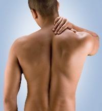
Herniated disc in the area of the cervical spine
In the case of a herniated disc in the area of the cervical spine, the gelatinous core usually swells out through the rear longitudinal ligament into the spinal canal or nerve canal. This leads to the nerves exiting through the lateral vertebrae being squeezed, which can lead to considerable back pain or neck pain radiating into the arm, shoulder or between the shoulder blades; paralysis with numbness is also possible. A very large herniated disc can even damage the spinal cord, requiring immediate emergency surgery.
Careful diagnosis – basic requirement for successful treatment
In order to be able to treat you successfully, your pain must be traced back to the segment to be operated on. A careful clinical examination in combination with imaging methods such as computer or magnetic resonance imaging is therefore the be-all and end-all for successful therapy of your disc disease.
Common complaints
- Neck pain that extends to the head and/or shoulder region
- Pain, dysesthesias, or weakness extending from the neck to the arms, backs of the hands, and/or fingers
- Shoulder pain with sensory disturbances in the deltoid area and/or weakness or paralysis when lifting the arm sideways.
The individual symptoms depend primarily on which section of the cervical spine is affected or whether and to what extent neighboring nerves are squeezed by the herniated disc.
What is a herniated disc?
The intervertebral discs are elastic cartilaginous discs about 5 mm thick and lie between the vertebral bodies of the spine. Its main function is to act as a "shock absorber" to absorb shock and vibration. In the center of the intervertebral disc is a gelatinous core surrounded by a solid ring of fibrous cartilage and connective tissue (fibrous ring).
In the case of a herniated disc, parts of the gelatinous core escape through tears in the fibrous ring into the intervertebral bodies or the spinal canal. As a result, there may be bruising and narrowing (compression) of the spinal cord or nerves exiting the spinal cord (spinal nerves); in this case, the acute back pain is accompanied by neurological symptoms in the supply area of the affected nerve root.
What causes a herniated disc in the cervical spine?
The starting point is usually degenerative changes, as a result of which the affected intervertebral disc tissue loses its elasticity and strength. Chronic poor posture or overloading due to professional or sporting demands can mean that even in younger people the natural compensation mechanisms are exceeded and the strained intervertebral disc can no longer withstand the high pressure. The consequences then range from a bulging of the gelatinous core into the fibrous ring to a veritable herniated disc. As a result, local back pain or neck pain, and more rarely radiating pain in the arm or hand, can occur.
In rare cases, an accidental injury can also promote a herniated disc in the cervical spine area.
A herniated disc in the cervical spine accounts for about eight percent of herniated discs and is therefore relatively rare compared to a herniated disc in the lumbar spine (in about 90 percent of cases).
How is it treated?
- Conservative treatment
A herniated disc of the cervical spine without paralysis is usually initially treated conservatively with pain-relieving medication and rest; Accompanying measures such as heat or electrotherapy as well as physiotherapy under physiotherapeutic guidance can provide relief.
In stubborn cases, injections of local anesthetics and cortisone preparations next to the constricted nerve root (periradicular infiltration) or next to the spinal cord (peridural infiltration) also help in the short term. In the case of severe neurological deficits, inpatient treatment with infusions containing cortisone to reduce swelling and inflammation, as well as painkillers, may also be necessary.
If the symptoms do not improve significantly within 6 - 8 weeks, an operative therapy should be considered!
- Surgical treatment
Another special focus in the APEX Spine Center is the percutaneous nucleotomy (intervertebral disc surgery) in the area of the cervical spine. This revolutionary surgical technique is almost exclusively performed by Dr. Schubert performed. In contrast to other surgical methods, the complete intervertebral disc is not removed and replaced by a placeholder (cage, bone block or prosthesis), as is usually the case, but only the herniated disc is removed under local anesthesia. The natural mobility and stability of the cervical spine is thus retained.
Ultimately, this surgical method is analogous to disc surgery in the lumbar spine area, where only the herniated disc is removed and not the entire disc is replaced by an artificial placeholder.